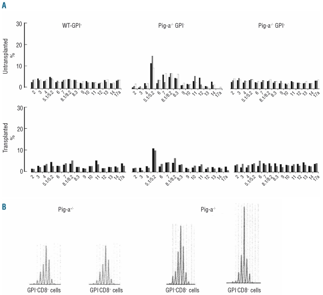
Figure 4.
GPI-deficient cells show T cell clonality. (A) Usage of TCR β variable region by CD8 T cells was analyzed in three WT-GPI+ from Pig-a+/+, three Pig-a−/− GPI−, two T-WT-GPI+, two TR-GPI−, and two TR-GPI+ bone marrow samples. The average of Vβ 5.1/5.2 in the GPI−CD8 T cells from Pig-a−/− mice and TR-GPI− mice was 22–23±5% compared to an average of 8–9.1±0.5% in GPI+ CD8 T cells and TR-GPI+ CD8 T cells. (B) Vβ skewing was confirmed by CDR3 analysis using specific primers for Vβ5.1 on sorted GPI−CD8+ and GPI+CD8+ cells from splenocytes of, respectively, Pig-a+/− and Pig-a−/0. The Vβ5.1 displayed 47% of total peak area in GPI−CD8+ T cells compared to 38% GPI+CD8+ from the Pig-a−/0 animal.

