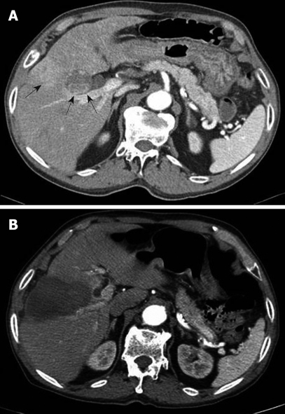
Figure 1.
Computed tomography (CT) scan of the patient. A: CT scan in an arterial phase demonstrates a 3.5 cm hepatocellular carcinoma lesion (arrows) at segment IV of the liver adjacent to the gallbladder, posterior to the lesion where a previous percutaneous ethanol injection has been performed; B: CT scan obtained after radiofrequency thermal ablation reveals complete ablation for the hepatocellular carcinoma without apparent complications such as a gallbladder injury on the immediate follow-up.

