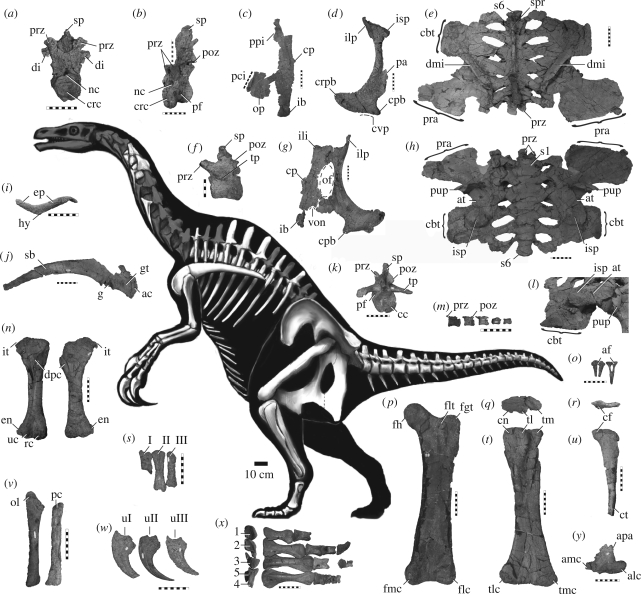
Figure 1.
Skeletal reconstruction (preserved shown in white) and representative elements of N. graffami (UMNH VP 16420): (a) cranial dorsal vertebra, cranial view; (b) mid-dorsal vertebra, craniolateral view; (c) left ischium, lateral view; (d) left pubis, lateral view; (e) dorsoventrally crushed iliosacrum, dorsal view; (f) proximal caudal vertebra, left lateral view; (g) right ischium/pubis, lateral view; (h) iliosacrum, ventral view; (i) furcula, cranial view; (j) right scapulacoracoid, lateral view; (k) proximal caudal vertebra, caudal view; (l) right iliosacrum, oblique ventrolateral view, showing crushed peduncles, acetabulum, brevis fossa and cubic tuberosity of postacetabular process; (m) four distalmost caudal vertebrae, lateral view; (n) left and right humeri, cranial view; (o) chevrons, cranial view; (p) left femur, cranial view; left tibia (q) proximal and (t) caudal views; left fibula (r) proximal and (u) lateral views; (s) left metacarpus, dorsal view; (v) antebrachium; (w) manual unguals, lateral view; (x) proximal (left) and dorsal (right) views of pes (missing PI–II, PIII–II, PIII–IV and PIV–V); and (y) left astragalus, cranial view. For abbreviations, see electronic supplementary material. Scale bars: 10 cm (a–e, g–y); 5 cm (f). Skeletal drawing modified from Victor O. Leshyk, copyright 2007.

