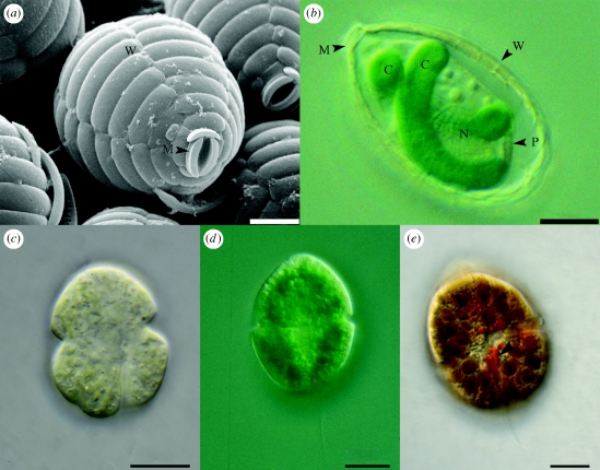
Figure 1.
Paulinella chromatophora cells. (a) Scanning electron microscopic image. (b) Light microscopic image (differential interference contrast). C, chromatophore; M, mouth opening; N, nucleus; P, plasma membrane and W, cell wall composed of silica scales. Light microscopic images of dinoflagellates with unusual plastids. (c) L. chlorophorum, (d) G. aeruginosum, and (e) K. foliaceum, arrowhead highlights the eyespot. Images kindly provided by Barbara Surek (c,e) and Karl-Heinz Linne von Berg (d). Scale bar, (a,b) 5 µm; (c–e) 10 µm.

