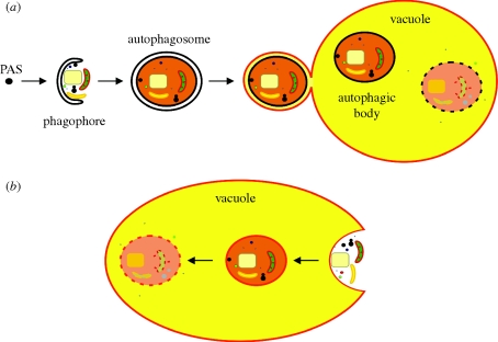
Figure 1.
Macro- and microautophagy in yeast. (a) Schematic representation of macroautophagy in baker's yeast. Upon induction of (nitrogen) starvation, the PAS (pre-autophagosomal structure or phagophore assembly site) incorporates membrane material and grows out to become the double membrane-layered phagophore that randomly sequesters cytoplasmic components (proteins and organelles). After complete engulfment of the cytoplasmic material, an autophagosome is formed. Subsequently, the outer membrane of the autophagosome fuses with the vacuolar membrane. As a result, this membrane obtains vacuolar characteristics (red colour) and ultimately becomes part of the vacuolar membrane, thereby increasing the size of the organelle. Additionally, a single membrane-bound autophagic body enters the vacuole, where it will become degraded by vacuolar hydrolases. (b) Schematic representation of microautophagy in baker's yeast. During microautophagy, the membrane of the vacuole engulfs a portion of the cytoplasm (including organelles). As a result, a vesicle with a single membrane originating from the vacuolar membrane (red colour) is formed inside the vacuole. This contrasts to macroautophagy, where the membrane of the autophagic body originates from the autophagosome (black colour). After complete engulfment, the vesicle and its contents are degraded by vacuolar hydrolases. Consequently, during microautophagy, the size of the vacuolar membrane is reduced. During selective forms of autophagy, mechanisms similar to macro- and microautophagy are used to selectively package peroxisomes, mitochondria, endoplasmic reticulum, ribosomes and portions of the nucleus (see text for details).

