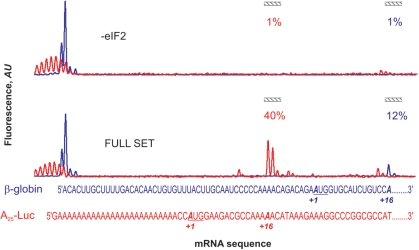
Figure 9.
Detection of the 48S complex assembly sites in the mixture of natural β-globin mRNA (FAM fluorescence, blue line) and synthetic A25-Luc mRNA (JOE fluorescence, red line). Both fluorescence readouts from FAM and JOE are normalized by the integral fluorescence intensity of the corresponding wavelength. The aligned β-globin and A25-Luc mRNA sequences are shown in the bottom. Complex-specific electrophoregram zones are indicated, and relative yield of the 48S complex assembled on the corresponding mRNA is shown.

