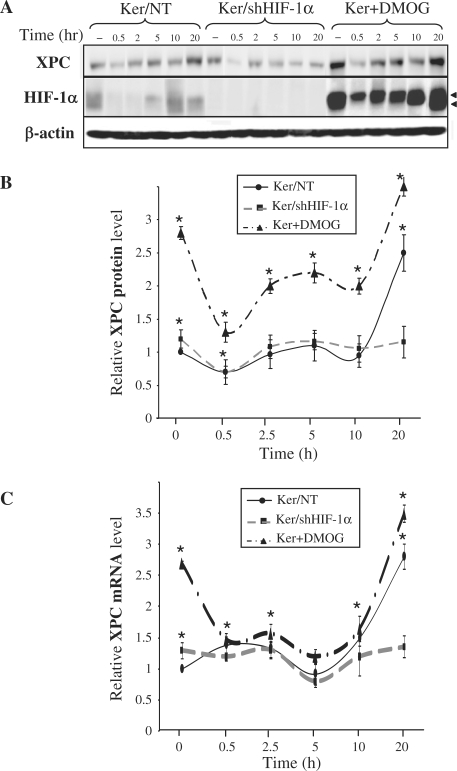
Figure 1.
Effect of UVB on XPC expression. Keratinocytes were harvested at the indicated time points after irradiation. (A) Total protein extracts were assessed for the presence of the XPC and HIF-1α protein by western blotting. β-actin was used as a loading control. Arrowheads indicate the two HIF-1α forms [see also ref. (14)]. (B) The band intensities of XPC proteins were quantified densitometrically. (C) The relative level of XPC mRNA was determined by qRT-PCR. The results are expressed as the mean ± SD of three independent experiments. –, no UVB exposure; Ker/NT, non-transduced keratinocytes; Ker/shHIF-1α, keratinocytes transduced with shHIF-1α; Ker + DMOG, keratinocytes treated with dimethyloxaloylglycine. *P < 0.05 for cells at the indicated time point versus non-transduced cells prior to irradiation.

