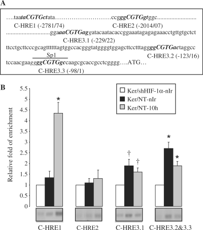
Figure 2.
Differential binding of HIF-1α on XPC promoter in non-irradiated and irradiated keratinocytes. (A) Five-nucleotides sequences matching the consensus HRE [(A/G)CGTG, marked in bold] are present in the 3 kb upstream region of human XPC gene, here referred to as C-HRE1, -HRE2, -HRE3.1, -HRE3.2 and -HRE3.3. The nucleotide sequences were numbered in relation to the translational start codon, ATG. The upperlined sequence represents the Sp1 binding site sharing a sequence in common with C-HRE3.3. (B) Ten hours after UVB-exposure, irradiated and non-irradiated keratinocytes were subjected to ChIP assay using an anti-HIF-1α antibody. Bands indicate PCR products using primers that span the indicated HREs. The sets of primers used are indicated in Supplementary Table S2. The relative levels of corresponding precipitated HRE fragments following ChIP were quantified by qRT-PCR. PCR amplification in shHIF-1α-transduced cells detected a minimal level of HREs which was normalized to one for each experiment. Ker/NT-10 h, non-transduced keratinocytes harvested 10 h after UVB exposure; nIr, non-irradiated keratinocytes. The results are expressed as the mean ± SD of three independent experiments. *P < 0.01 and †P < 0.05 for each HRE versus shHIF-1α−transduced keratinocytes.

