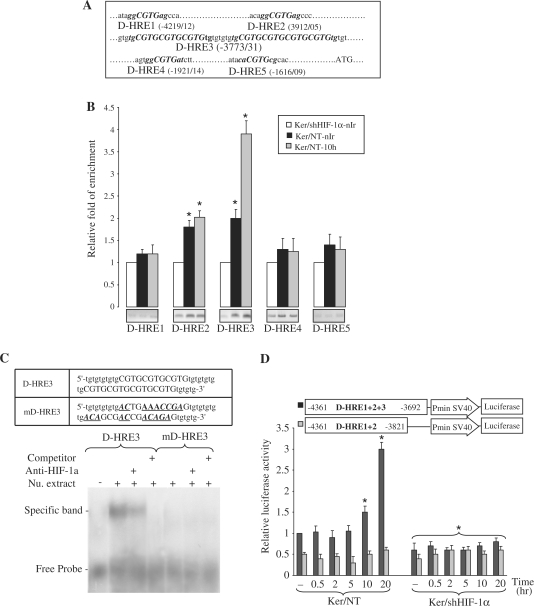
Figure 5.
Binding of HIF-1α to the XPD promoter in non-irradiated and irradiated keratinocytes. (A) Eleven-nucleotides sequences matching the consensus HRE (marked in bold) are present in the 4 kb upstream region of the human XPD gene, here referred to as D-HRE1 to 5, in which D-HRE3 includes seven overlapping HREs. (B) Ten hours after UVB-exposure, irradiated and non-irradiated keratinocytes were subjected to ChIP analysis using an anti-HIF-1α antibody. Bands indicate PCR products amplified using primers that span the indicated HREs. The relative levels of corresponding precipitated HRE fragments following ChIP were quantified by qRT-PCR. The results are expressed as the mean±SD of three independent experiments. *P < 0.01 for each HRE versus shHIF-1α-transduced keratinocytes. (C) EMSA analysis of the XPD promoter using HRE mutant probe and anti-HIF-1α antibody. The sequences of the oligonucleotide probes are presented in the top panel. D-HRE3 is the wild-type probe and mD-HRE3 is the probe mutated in the HIF-1α-binding sites. Mutated bases are indicated with bold, italics and underlining. (D) Relative luciferase activities were measured in NT or shHIF-1α-transduced keratinocytes transfected with luciferase reporter constructs containing either the D-HRE1 + 2 + 3 or the D-HRE1 + 2 at the indicated time points after UVB irradiation. For each reporter, the mean luciferase activity is shown (±SD, n = 3) relative to the activity in the NT cells prior to irradiation. *P < 0.05 for cells at the indicated time point versus NT cells prior to irradiation.

