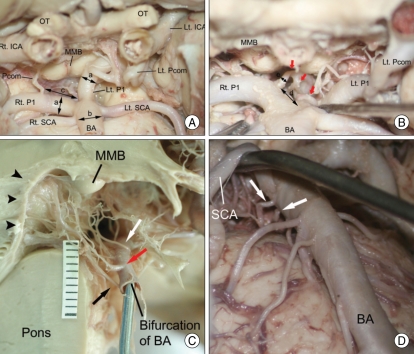
Fig. 1.
Photographs showing the anatomical structures in the interpeduncular fossa. The measured vascular structures are visible on the anteroinferior view of the interpeducular fossa (A and B). On the midsagittal section of the brain stem (C), thalamoperforating arteries arise from the superior (white arrow), posterosuperior (red arrow) and posterior surfaces (black arrow) of the P1 segment and enter the midbrain through the posterior perforated substance (arrowheads). There are visible two perforating arteries (white arrows) arising from the ventral surface of BA on the anteroinferior view of the interpeduncular fossa (D). a, diameter of the P1 segment; b, diameter of the BA; c, distance of the P1 segment; d, distance between the bifurcation of BA and the 1st origin of the thalamoperforating artery (red arrows); e, diameter of the thalamoperforating artery. BA : basilar artery, ICA : internal carotid artery, Lt : left, MMB : mamillary body, OT : optic nerve, Pcom : posterior communicating artery, P1 : P1 segment of posterior cerebral artery, Rt : right, SCA : superior cerebellar artery.

