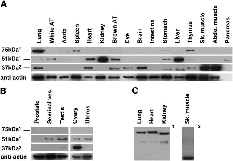
Fig. 4.
FADS3 distribution in rat tissues. The occurrence of FADS3 was determined in different rat tissues. SDS-PAGE (A and B) and native PAGE (C) were performed on protein lysates from somatic male tissues (A) and from male and female germinal tissues (B). Western blots were then assessed using rat anti-FADS3 antibodies (1anti-NtermFADS3 and 2anti-CtermFADS3). The intensity of band observed in abdominal and skeletal muscles was deliberately reduced by five because of the excessive signal. Experiments were reproduced on three rats; results exhibit only one case in point.

