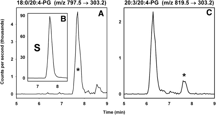
Fig. 3.
Examples of extracted ion chromatograms for selected PG lipids. In panel A, the peak corresponding to 18:0/20:4-PG is shown eluting at 7.8 min, along with panel B as an inset showing that the synthetic standard 17:0/20:4-PG (labeled ‘S’) elutes at the same time. In panel C, the peak representing a minor species is also shown eluting at 7.8 min. The peak at 6.3 min is most likely an AA-containing triglyceride (e.g., 12:0/18:3/20:4-TG), because it coelutes with species shown by MS/MS analysis to represent 10:0/20:4/22:6-TG. The asterisk in panels A and C indicates that the parent ions also yield [M−171]+ fragment ions in positive mode MS/MS studies.

