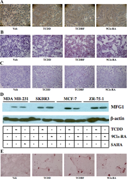
Figure 6.
Activation of AhR induces markers of breast epithelial cell differentiation. A, SKBR3 cells were seeded in six-well plates and treated with vehicle (Veh), TCDD (10 nm), TCDBF (10 nm), or 9-cisRA (1 μm) for 48 h. Cells were imaged by light microscopy. SKBR3 cells (B) and MCF-7 cells (C) were seeded in six-well plates and treated for 48 h as described above. Cells were fixed, stained with Oil Red O, counterstained with hematoxylin, and imaged by light microscopy. Red granules represent cellular lipid deposits. For A–C, a representative experiment (n =3 independent experiments each) is shown in each case. D, SKBR3, MDA MB-231, MCF-7, and ZR-75-1 cells were treated for 48 h with Veh (lanes 1, 4, 7, and 10), 10 nm TCDD (lanes 2, 5, 8, and 11), 500 nm SAHA (lane 3), or 1 μm 9-cisRA (lanes 6, 9, and 12). Cells were lysed and whole-cell extracts were prepared and used for SDS-PAGE and Western blotting using a rabbit antihuman antibody to MFG1. A mouse antihuman β-actin antibody was used as a control for total protein between different samples. Shown is a representative experiments (n = 4 independent experiments). E, LA7 cells were seeded in four-well chamber slides and treated with TCDD, TCDBF, or 9-cisRA as described above for 48 h. Cells were fixed, stained with Oil Red O, counterstained with hematoxylin, and imaged by light microscopy.

