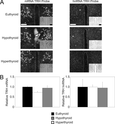
Figure 2.
TRH mRNA is expressed in the DMH. ISH using S35 radiolabeled probes. A, ISH was performed on brain slices from euthyroid, hypothyroid, and hyperthyroid WT mice using the TRH mRNA (left column) and TRH hnRNA (right column) probes. Magnification, ×10; scale bars per column, 100 μm. n = 4 mice for each probe under each condition. B, Quantification of ISH via pixel density. Data are presented as sample means relative to control euthyroid mice ± sem. Significance was tested within each probe via one-way ANOVA.

