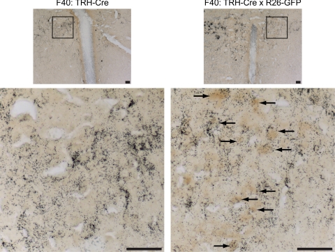
Figure 4.
GFP-positive neurons colocalize with neurons expressing TRH mRNA. Dual-label ISH/IHC for TRH mRNA using a S35 radiolabeled riboprobe (black grains) and immunostaining for GFP (dark brown) was performed in Founder 40 TRH-Cre (F40: TRH-Cre) and F40: TRH-Cre crossed to Rosa-26-GFP reporter (F40: TRH-Cre x R26-GFP) mice. Magnifications, ×10 (top) and ×40 (bottom) of the highlighted area of the PVH are shown; scale bars, 100 μm. Arrows identify neurons positive for both TRH mRNA and GFP immunostaining.

