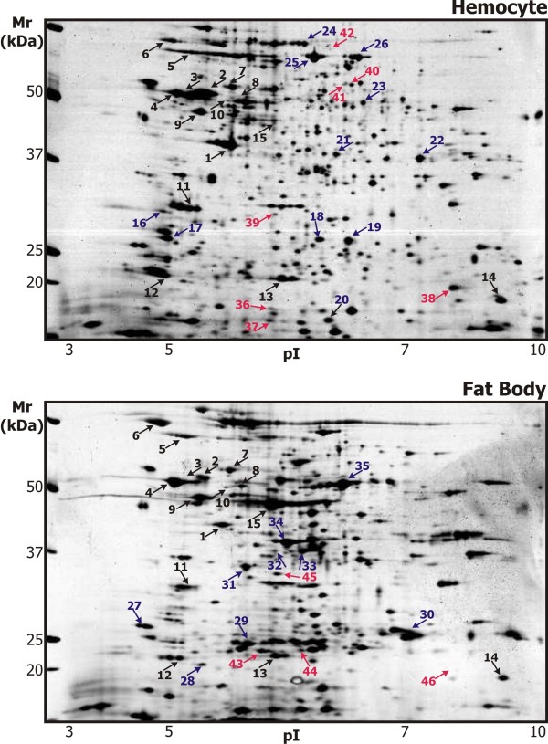
Figure 1.
Representative 2-DE maps of proteins of the S. bullata larvae hemocytes and fat bodies. Solubilized proteins from the S. Bullata hemocytes and fat bodies, 6 hours after the injection (induced) or the treatment with sterile entomological pin (non-induced), were focused on IPG strips pH 3-10 NL and separated in SDS-polyacrylamide gradient gels (8-16%). The gels were silver stained. The proteins identified in numbered spots are listed in Additional file 1. The proteins identified in both 2-D protein maps are in black; proteins identified either in the hemocytes or in the fat bodies are in blue, and the proteins changed after immune challenge are in red.

