
Figure 1.
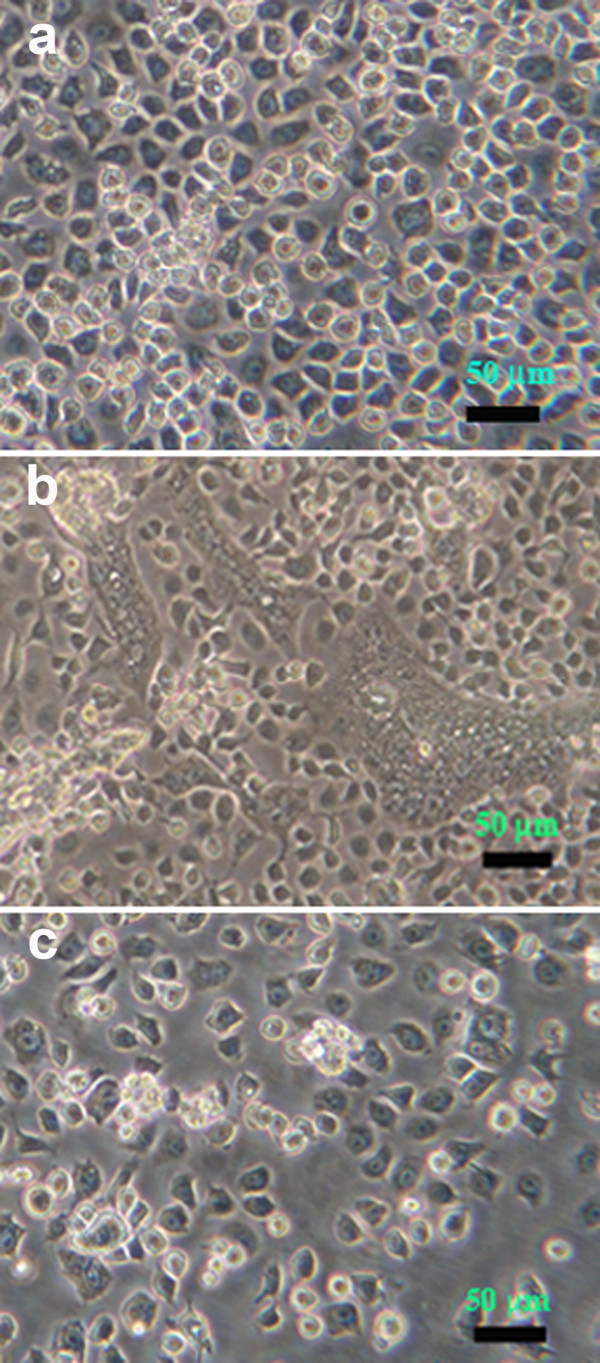
Phase contrast photomicrographs of C6/36 cells. (a) Naïve cells. (b) Cells with triple co-infections at passage 2 showing some cytopathology. (c) Cells with triple co-infections at passage 4 with morphology similar to that of naïve cells and of cells from higher passages.