
Figure 2.
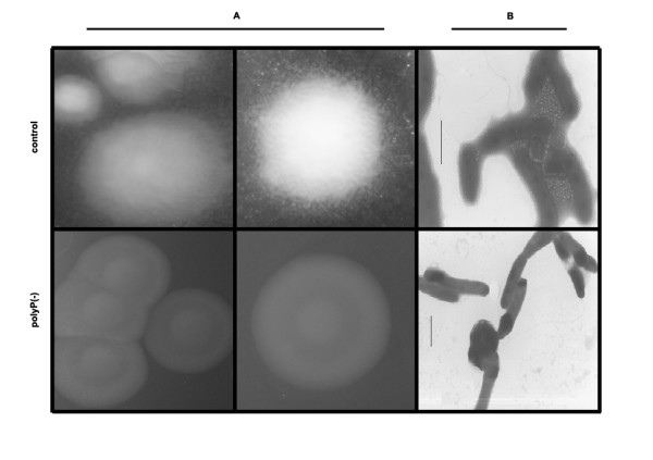
Colony morphology of polyP-deficient cells of Pseudomonas sp. B4. Pseudomonas sp. B4 polyP-deficient and control cells were grown in LB plates for 48 h and the colonies were photographed by using a magnifying glass (A). Unstained cells were analyzed by transmission electron microscopy (B).