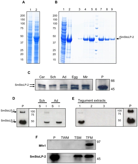
Figure 2. SDS-PAGE (4%–12%) analysis of cell extracts and fractions from E. coli (BL21DE3) transformed with the pDEST17-SmStoLP-2, and immunoblotting of protein extracts from S. mansoni stages and fractions using anti-rSmStoLP-2 polyclonal antibodies.
(A) Lanes 1 and 2 represent a clone before and after induction with 1 mM IPTG, respectively; (B) Inclusion bodies were extracted with urea and denatured protein was refolded by dilution before being purified through Ni2+-charged column chromatography. Lanes 1 and 2 show the soluble fraction after lysis and the inclusion bodies after solubilization with 8 M urea, respectively. Lanes 3–5 and 7–9 show the fractions of rSmStoLP-2-6xHIS-tag fusion protein eluted after Ni2+ chromatography, Lane 6, MW ladder (kDa); (C) Immunoblotting of S. mansoni extracts from different stages using anti-rSmStoLP-2 polyclonal antibodies (20 µg of protein were loaded in each lane). Cer – cercariae, Sch – 7-day-old schistosomula, Ad – adult worms, Egg – eggs, Mir – miracidia. (D) Western blot of soluble (S) and insoluble (I) protein extracts of 10-day-old schistosomula (Sch) and adult worms (Ad); (E) Detection of SmStoLP-2 in the tegument of S. mansoni adult worms, (1) proteins soluble in urea and thiourea, (2) proteins soluble in urea, thiourea, CHAPS and SB 3–10, (3) proteins soluble in 2% SDS. (F) Dual targeting of SmStoLP-2 to tegumental membranes and tegumental mitochondria, TWM, tegument extract without surface membranes, TSM, tegument enriched in surface membranes and TFM, tegument fraction enriched in mitochondria (20 µg of protein were loaded in each lane), Mfn-1 – is the Mitofusin-1 mitochondrial marker. Arrows indicate the rSmStoLP-2 and the most reactive bands of native SmStoLP-2 detected in each experiment. Positions of molecular mass standards (kDa) are indicated on the right or in the center. Positive control (P) contains 50–60 ng of rSmStoLP-2.

