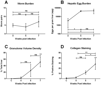
Figure 1. Parasite burden and granulofibrotic pathology.
Infected mice harboured a mean of 5 worm pairs (A). Schistosome eggs were first observed in the liver at 4 weeks p.i and hepatic egg burden increased significantly thereafter (1-Way ANOVA, p≤0.05) (B). Granuloma volume (C) and collagen staining for hepatic fibrosis (D) increased significantly from 4 weeks p.i, reaching 51% and 28% total liver volume at 7 weeks p.i, respectively (1-Way ANOVA, p≤0.01). Values represent mean values from 4 mice pooled for microarray analysis ±1SD.* p≤0.05, ** p≤0.01, ns = not significant compared with uninfected liver unless otherwise indicated.

