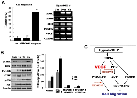
Figure 4. De-differentiated ATSCs evidenced active cell migration.
(A) Migration activity of de-ATSC was evaluated as a percentage of the spontaneous migration and related functional factors. The migration activity of the dedifferentiated ATSC in vitro, the cells were transferred to culture dishes containing low serum growth medium. The cultured cells were transferred into transwell membranes (8 µm pore size), coated on both sides with laminin. In the upper chamber, both of cells were preincubated in a CO2 incubator. For analysis, migrating cells on the lower surface were air-dried and counterstained with Harris hematoxylin and the numbers of cells on the lower surfaces were assessed. Ten x20 fields per insert were counted. (B) The migration activity of dedifferentiation of ATSCs was caused by ERK, JUNK, and P38 phosporylations. (C) Proposed molecular mechanism of cell migration after ATSCs reprogramming by DHP-d/Hypoxia. Datas presented are presented as mean ±SD; n>4. * p % 0.05, and ** p % 0.01, Student's t test.

