
Figure 4.
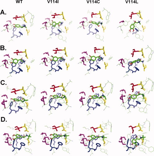
Comparison of the binding pockets for β2-AR agonist ligands in the wild-type (WT) and the three mutants V114I, V114C, and V114L (from left to right). Residues within 5 Å of the ligand are shown. Highlighted as sticks are the following entities: the ligands are shown in Cory-Pauling-Kolton coloring scheme. The residue at position 114 is shown in red. Residues D113 and N312 are colored in yellow, and W286, F289, and F290 are colored in blue and represented in sticks. S203, S204, and S207 are colored in magenta. (A) Epinephrine, (B) norepinephrine, (C) isoproterenol, and (D) Salbutamol.