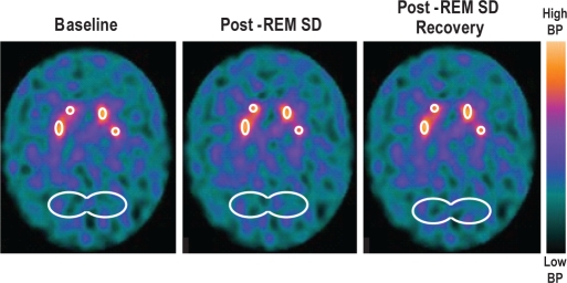
Figure 2.
Averaged brain images based on three consecutive slice regions of interest (ROIs) for [99mTc]TRODAT-1 were used to estimate the concentration of DAT in the striatum (right and left at baseline) post-SD, and post-sleep recovery. An elliptical ROI was placed on three consecutive slices in the occipital cortex, an area used for reference of non-specific DAT binding.

