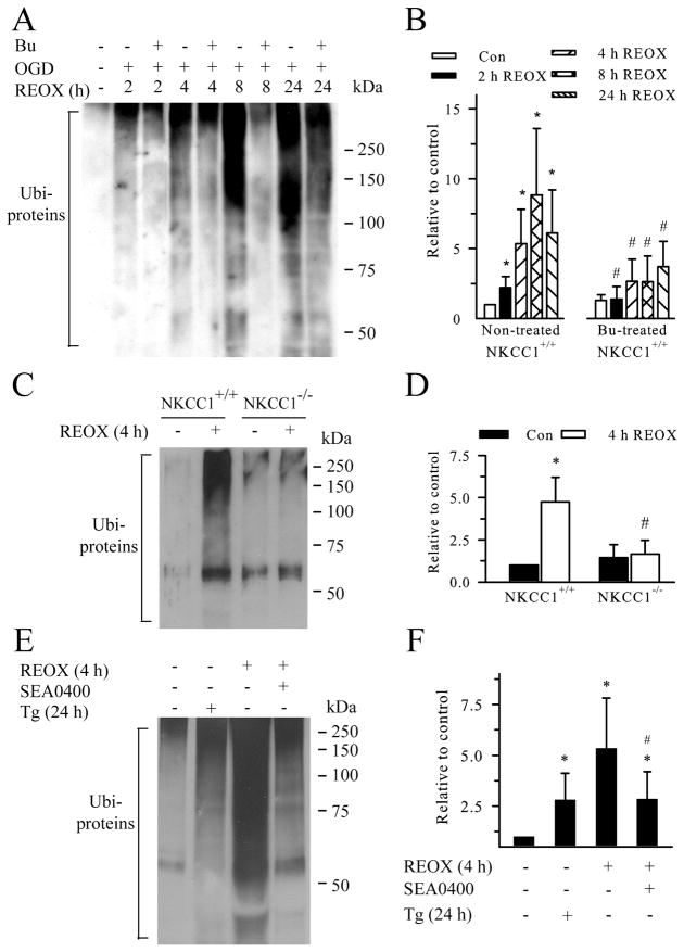
Figure 1. Inhibition of NKCC1/NCXrev attenuates protein aggregate formation following OGD/REOX.
A. Inhibition of NKCC1 activity by bumetanide attenuated formation of ubi-protein aggregates. TX-100-insoluble protein fractions were obtained from cellular lysates (500 μg protein) of normoxic controls and OGD/REOX-treated cultures. Protein smear ranged between 37–250 kDa in each lane as detected by a monoclonal anti-ubiquitin antibody. In the case of Bu treatment, 10 μM Bu was added 30 min prior to OGD and present in all subsequent incubations. B. Summary data. Ubi-proteins were quantified using average intensity of protein smear in each lane with ImageJ software and data were presented as relative change to control. Data are mean ± SD. n = 3–7. * p < 0.05 vs. normoxic control; # p < 0.05 vs. non-treated. C. Effects of NKCC1 gene ablation on formation of ubi-proteins. NKCC1−/− and NKCC1+/+ cultures from the same littermates were subjected to 2 h OGD and 4 h REOX in parallel. D. Summary data. Data are mean ± SD. n = 4. * p < 0.05 vs. NKCC1+/+ normoxic control; # p < 0.05 vs. NKCC1+/+ 4 h REOX. E. Effects of inhibition of NCXrev activity or SERCA on formation of ubi-proteins. NCXrev inhibitor SEA0400 (1 μM) was present at 0–4 h REOX or 0.1 μM Tg was present for 24 h. F. Summary data. Data are mean ± SD. n = 3–4. * p < 0.05 vs. normoxic control; # p < 0.05 vs. 4 h REOX. Statistical significance was determined by paired student’s t test between the two groups.

