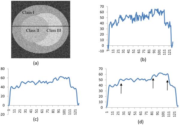
Fig.4.
Anisotropic filter processing for a simulated noisy image. (a) is the original image labeled with three classes (Class I, II, III). The image is corrupted with noise. (b) is the signal profile along the labeled center line on Image (a). (c) and (d) are the signal profiles at Scale 5 and Scale 7 by diffusion filtering, respectively. The noise is removed and the line is smoothed. Furthermore, the edges are enhanced as shown by the arrows in (d).

