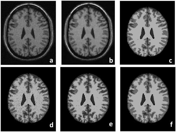
Fig.7.
Classification results of brain MR images. The original MR image (a) is smoothed after the anisotropic filter processing (b). (c) is the ground truth of the classification. (d), (e), and (f) are the classification results using the FCM, MFCM, and MsFCM methods, respectively. Compared to the ground truth, the MsFCM performs better than the other two methods. The image was obtained from the McGill brain database with 9% noise and 20% intensity inhomogeneity.

