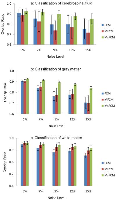
Fig.8.
Overlap ratios of the classification results for the three methods, i.e. FCM, MFCM, and MsFCM. The images were obtained from the McGill brain database with 9% noise and 20% inhomogeneity. Each bar represent 5 classifications from 5 images, Mean and standard deviation are plotted. The MsFCM achieved higher overlap ratios compared to the other two methods.

