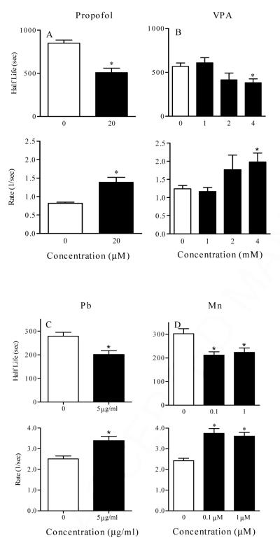
Figure 5.
Analysis of half-life and rate (K) of Ca2+ wave initiated in CCF-STTG1 cells treated for 24 hr with propofol (0, 20 μM) (top, left panel), valproic acid (0-4 mM) (top, right panel), lead (Pb) (0, 5 ug/ml) (bottom, left panel), and Manganese (Mn) ( 0- 1 μM ) (bottom, right panel). In each experiment, cells were loaded with fluo4 for 1 h at 37° C and then washed with serum free medium. The wave was then generated by addition of 10% FBS and cells were imaged for approximately 30 min at 5 seconds interval. Data represented was collected from at least 8 images, 15 to 30 cells per image, per treatment. Asterisk represents significant difference from control at p < 0.05.

