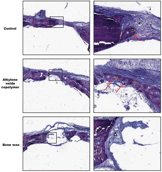
FIGURE 4.
Histological analysis of bone healing in calvarial defects. Representative images of 5-μm thick coronal sections from the center of the defect at 3 weeks after surgery. Toluidine blue staining demonstrates mineralized bone (blue stars), osteoblasts (red arrows), and osteoid (green arrowheads) in the control and alkylene oxide copolymer-treated defects, with mostly fibrous tissue observed at the site of the bone wax treated defects (white triangle). Both low (× 2, left) and high magnification (× 10, right) are shown.

