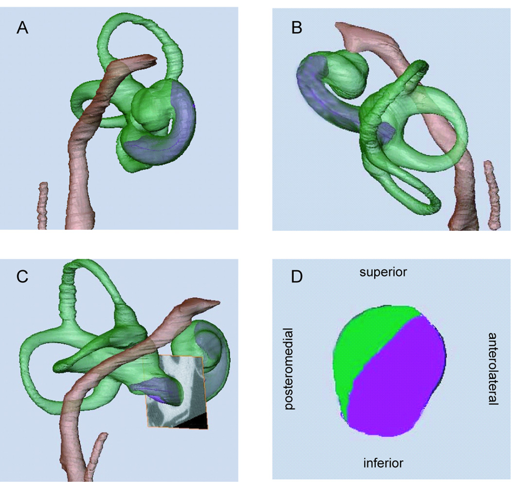
Figure 1.
View of segmented structures of right ear from anterior (A), superior (B), and lateral (C). A cross-section of the cochlea is shown in D, with the scala tympani shown in purple and the scala vestibuli shown in green. This cross-section corresponds to the plane shown in C, just anterior to the round window.

