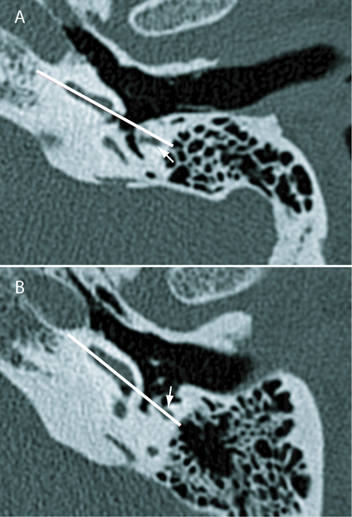
Figure 5.
Relationship of the plane of the basal turn of the cochlea to the facial nerve. These axial CT scans (done at an isotropic resolution of 100 microns) indicate rotation of the cochlea in the axial plane, with the apex facing more anteriorly (specimen shown in panel A) and facing more laterally (specimen shown in panel B). The white line is determined by the posteromedial face of the basal half of the basal turn, approximately representing the scala tympani. This indicates the approximate optimal vector of insertion coaxial with the centerline of the basal turn. White arrows represent the facial nerve. The line of insertion passes lateral to the facial nerve in the specimen shown in panel A but directly through it in the specimen shown in panel B.

