Fig. 1.
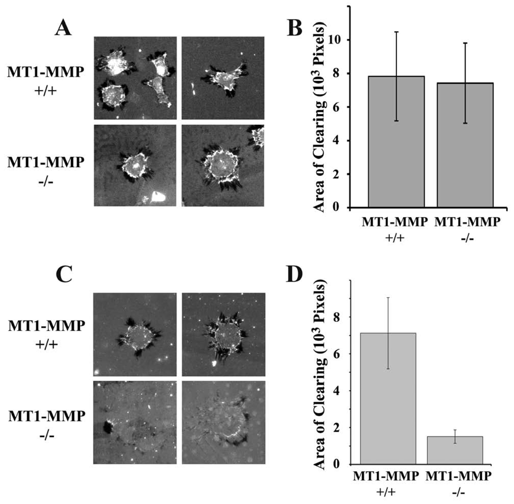
ECM remodeling by MT1-MMP-deficient (−/−) and expressing (+/+) fibroblasts. The substratum consisted of fluorescently-labeled fibronectin layered on top of type I collagen. (A) Representative cells are shown. Darkened or black areas represent “areas of clearing” or “AOCs”, which are measured as evidence of ECM remodeling. (B) Quantification of the total AOCs for individual MT1-MMP(−/−) or (+/+) cells. AOCs were measured using image J software (mean ± SEM). (C) MT1-MMP-deficient and wild type skin fibroblasts were plated on fibronectin layered over fluorescein-labeled type 1 collagen and allowed to remodel the ECM for 3 h prior to fixation. Representative cells are shown. Many MT1-MMP-defiicent cells showed no evidence of remodeling. (D) Quantification of the total AOCs for wild type and MT1-MMP-deficient cells (mean ± SEM).

