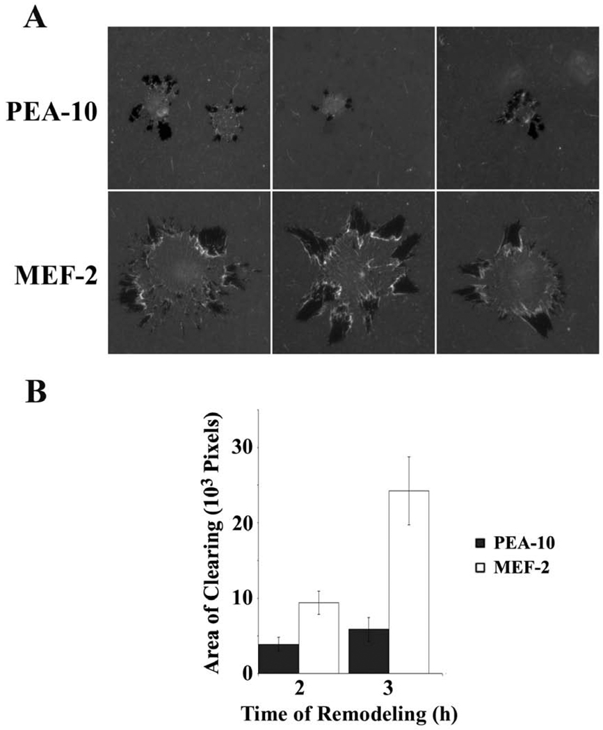
Fig. 2.
ECM remodeling by LRP1-deficient MEF-2 cells and LRP1-expressing PEA-10 cells. The substratum consisted of fluorescently-labeled fibronectin layered on top of type I collagen. (A) Representative cells are shown. Darkened or black areas represent AOCs. (B) Quantification of AOCs for individual PEA-10 and MEF-2 cells 2h and 3h after plating. AOCs were measured using image J software (mean ± SEM).

