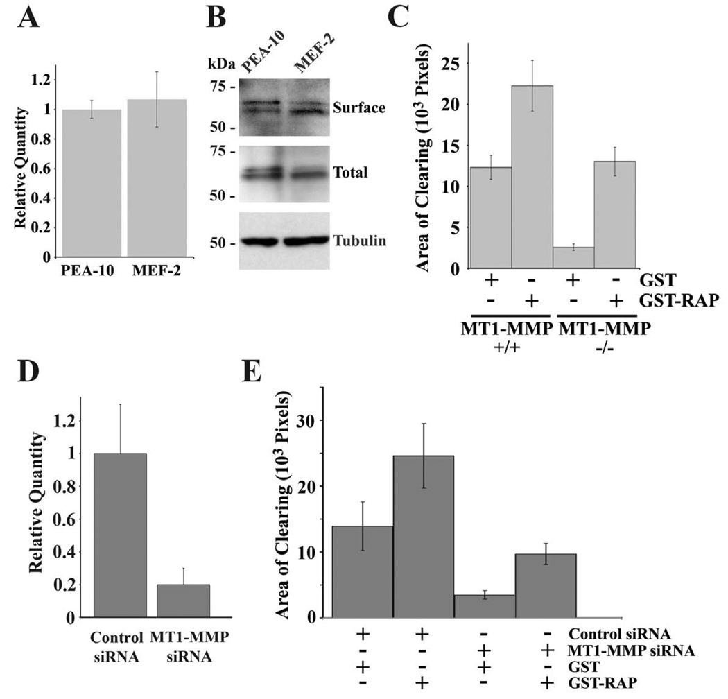
Fig. 6.
LRP1 regulates ECM remodeling in the presence and absence of MT1-MMP. (A) Total RNA was isolated from PEA-10 and MEF-2 cells and analyzed by qPCR using specific primers for MT1-MMP. (B) Equal amounts of cellular protein from detergentsoluble cell extracts of PEA-10 and MEF-2, which were labeled with biotin, were subjected to affinity precipitation. The precipitates were analyzed by immunoblot analysis to determine “surface” MT1-MMP. Whole cell extracts also were subjected to immunoblot analysis to detect “total” MT1-MMP and tubulin, as a loading control. (C) MT1-MMP-deficient and wild-type skin fibroblasts were cultured in the presence of GST-RAP (0.2 µM) or GST for 72 h. Cells were plated on fibronectin layered over fluoresceinlabeled type 1 collagen and allowed to remodel the ECM for 3 h prior to fixation. AOCs were measured using image J software (mean ± SEM). (D) PEA-10 cells were co-transfected to express RFP and with MT1-MMP-specific or non-targeting control siRNA. Total RNA was isolated from these cells and analyzed by qPCR using specific primers for MT1-MMP. (E) RFP-transfected PEA-10 cells in which MT1-MMP was silenced or not were cultured in the presence of GST-RAP (0.2 µM) or GST for 72 h. The cells were plated on fibronectin layered over fluorescein-labeled type 1 collagen. Transfected cells were allowed to remodel the ECM for 3 h prior to fixation. AOCs of RFP-positive cells were measured using image J software (mean ± SEM).

