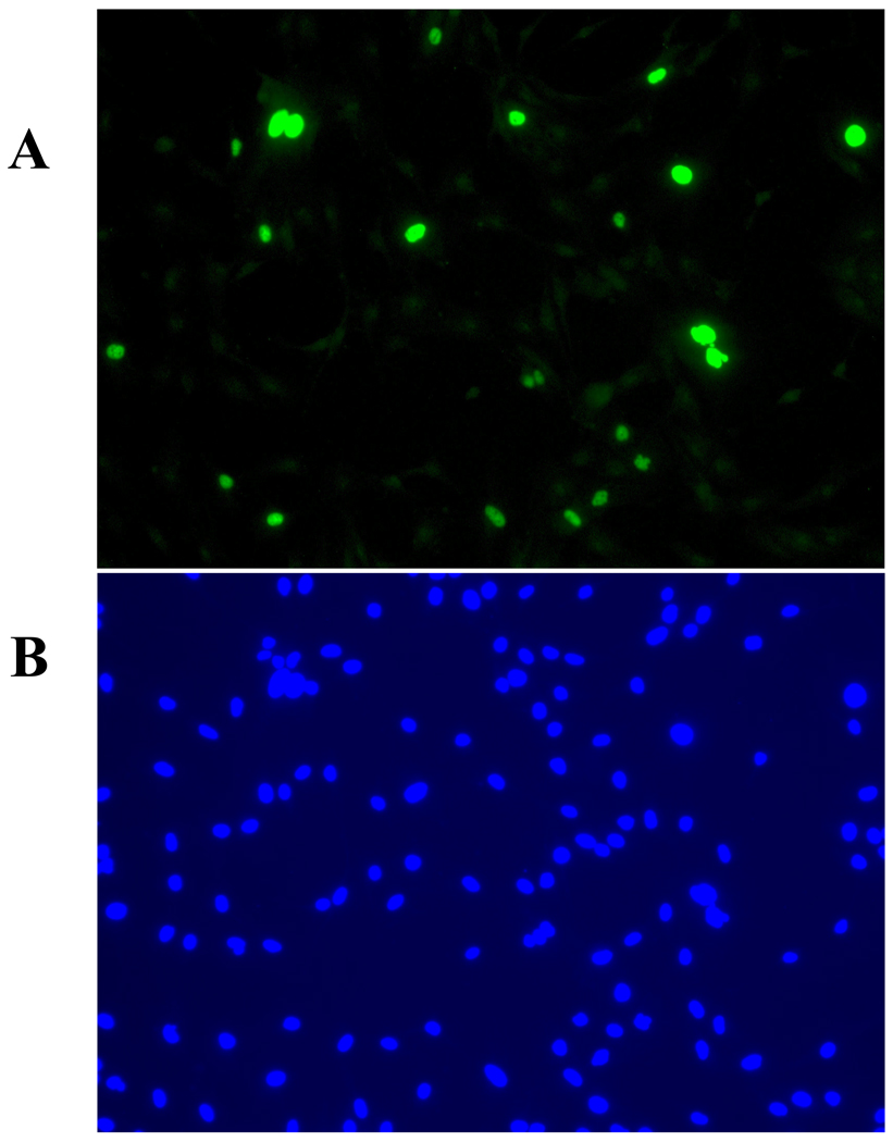
Fig. 1. Detection of HRPTEC BKV infection by immunofluorescence. HRPTEC are either stained with anti-T-Ag antibodies (A), or stained with 4’,6-diamidino-2-phenylindole, dilactate (DAPI) (B).
HRPTEC were incubated with BKV (MOI 0.5 FFU/cell). After 72 hours fresh medium was added and incubated for another 48 hours. After incubation, cells were fixed and blocked. Then cells were incubated with primary antibody {5 µl of PA 416 (Calbiochem, San Diego, CA) against 1 ml of TTBS with 1 % FBS} and second antibody{1:200 dilutied Alexa flour™ 488 goat anti-mouse IgG (H+L) (Molecular Probes, Eugene, OR) against TTBS with 1 % FBS}. To stain nuclei, cells were incubated with DAPI (300 nM) (Molecular Probes, Eugene, OR) for 5 minutes and washed three times with PBS. Cells were observed by fluorescent microscope (Nikon Eclipse E600) with 20× objective lens and images were captured by SPOT® version 4.0.9 (Diagnostic instruments, Scotland, UK).

