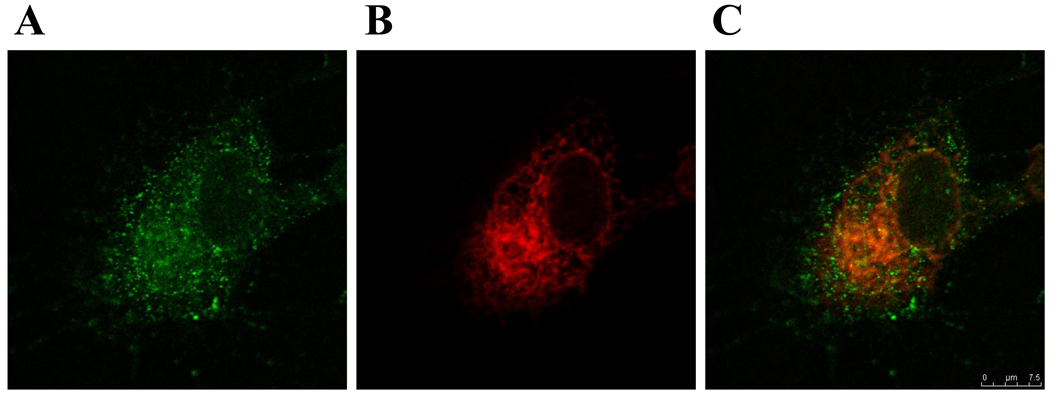
Fig. 2. Localization of labeled BKV particles in HRPTEC. (A) Fluorescence of Alexa Fluor 488 labeled BKV particles in HRPTEC, (B) Staining for Endoplasmic reticulum (ER) in HRPTEC, (C) Co-localization of purified and labeled BKV with ER marker.
HRPTEC were incubated with purified and labeled BKV for 6 hours. After incubation, cells were fixed and blocked. Then cells were incubated with primary antibody {1: 100 dilution of PDI (Abcam, Cambridge, MA) as ER marker against TTBS with 1 % FBS} and second antibody {1:200 dilutied Alexa flour™ 680 goat anti-mouse IgG (H+L) (Molecular Probes, Eugene, OR) in TTBS with 1 % FBS}. Cells were analyzed by confocal microscope (Leica TCS SP5) with 63× objective lens and images were captured by Leica application suite advanced fluorescence. Line is 10 µm.

