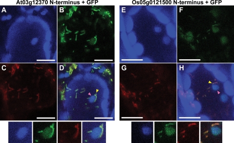
Figure 4.
Subcellular localization in Arabidopsis leaf epidermal cells of GFP fusion proteins having the N-terminal portion of the second chloroplast-like RPL10 copy from Arabidopsis (A–D) and rice (E–H). (A and E) Chloroplast autofluorescence. Note that a number of large particles that did not coincide with the GFP image represent autofluorescence from mesophyll chloroplasts that occur below the epidermal cells. (B and F) GFP fluorescence; (C and G) mt-DsRed fluorescence as a control of mitochondrial targeting; (D and H) merger of the three images (chloroplast autofluorescence, GFP fluorescence, and mt-DsRed fluorescence) (A–C and E–G, respectively). Mitochondria and chloroplasts are indicated by yellow and pink arrowheads, respectively. Scale bar = 10 µm. An enlarged portion of the same small subportion of each image is shown below the set of four full images.

