Fig. 4.
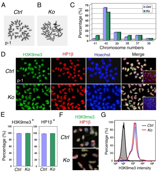
Surviving Dicer-deficient NSCs have normal karyotype and heterochromatin protein expression. (A,B) Karyotype analyses (metaphase spreads) of surviving Dicer-deficient (Ko) and control (Ctrl) NSCs collected from p-1. (C) The percentage of chromosome numbers among counted cells. More than 54% Dicer KO NSCs displayed 40 chromosomes, which is similar to the controls (63%); n=33 cells for either Ctrl or KO NSCs. (D) Immunohistochemistry for heterochromatin proteins in control and Dicer- deficient NSCs (p-1). Both H3K9me3 and HP1β were detected and coexpressed in Dicer-deficient NSCs. The insets in merged images show immunostaining without the primary antibodies for H3K9me3 and HP1β. (E) Almost 100% of control and Dicer-deficient NSCs were H3K9me3- and HP1β-positive. n>250 cells; three Dicer KO and three control animals were used for statistical analyses. (F) H3K9me3- and HP1β-expressing Dicer- deficient NSCs had elongated nuclei. (G) The intensity of H3K9me3 expression levels was similar between the control (blue line) and KO (red line) NSCs, as detected by the flow cytometric analysis. The gray area indicates an isotype-negative control. The representative result shown here is from three independent experiments.

