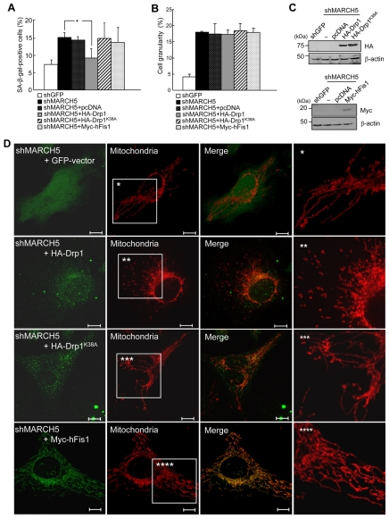
Fig. 7.
Ectopic expressions of Drp1 diminish the level of SA-β-Gal positivity induced by MARCH5 depletion. Hemagglutinin (HA)-Drp1, HA-Drp1K38A and Myc-hFis1 expression vectors were introduced into the MARCH5-depleted cells on day 2. After 2 days, cells were analyzed for SA-β-Gal positivity, cellular granularity and mitochondrial morphology. (A) Quantification of SA-β-Gal-positive cells. Approximately 300 cells were counted in several fields and data represent the average of four independent experiments. **P<0.01 vs MARCH5 shRNA by Student's t-test. (B) Cellular granularity was evaluated by analyzing the side-light scatter in flow cytometry. (C) The ectopic expression levels of HA-Drp1, HA-Drp1K38A and Myc-hFis1 were determined by immunoblotting. (D) Mitochondria were analyzed by confocal microscopy after staining with MitoTracker Red. Boxed areas are magnified on the right. Scale bars: 10 μm.

