Figure 3.
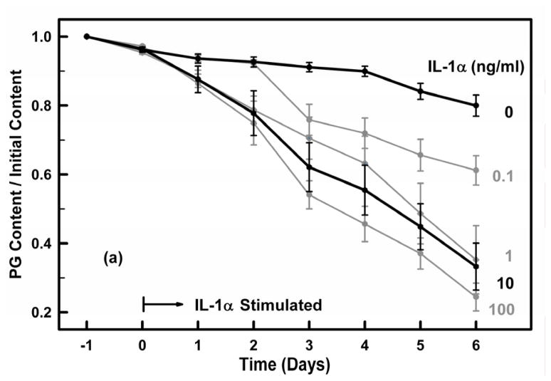
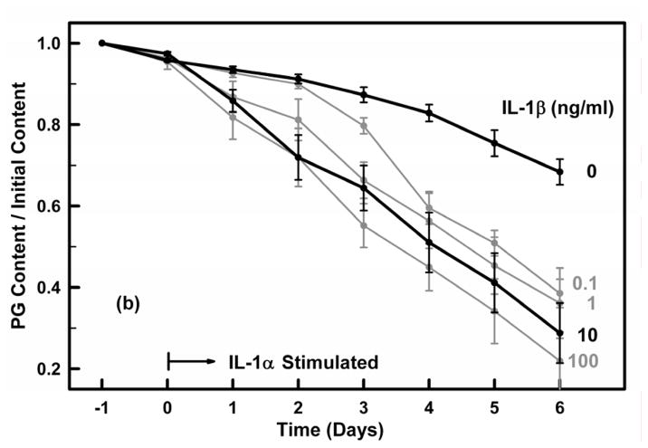
Figures 3a and 3b. Cartilage explants were incubated for 24 hours in IL-1 free media (days −1 to 0) and then were exposed on day 0 to (a) IL-1α and (b) IL-1β at concentrations of 0, 0.1, 1, 10 and 100 ng/ml for six days (days 1–6). The PG content remaining in the explant on each day was determined from the PG released to the media on each day and the PG remaining in the explant on day 6. Media and IL-1 were exchanged on each day. All groups had similar PG contents at the time of IL-1 administration on day 0. The 0 and 10 ng/ml concentrations are highlighted. Data are means ± SEM for n=5 explants.


