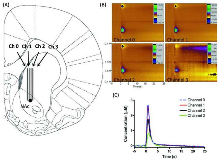
Figure 2.
Simultaneous in vivo dopamine detection. (A) Histological placement of MEA as confirmed by lesioning with a tungsten microelectrode post-experimentation (marked with a black circle). (B) Simultaneously collected color plots of electrically stimulated dopamine release. Stimulation parameters: 60 Hz, 40 pulses (± 300 μA, 2 ms per pulse). (C) Dopamine oxidation current vs. time traces at 4 channels. Stimulation delivered at t = 0 s.

