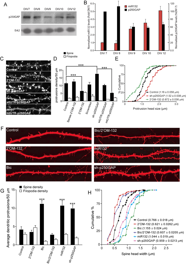
Fig. 2. miR132 regulates spine formation by repressing p250GAP.
(A) Developmentally timed hippocampal cultures were analyzed by Western blot for expression of p250GAP. (B) p250GAP expression was normalized to Erk2 levels and plotted with normalized mature-miR132 levels. (C–E) Hippocampal neurons DIV 7 were transfected with EYFP-γ actin and either empty vector (Control), Sense-2’OM-132 (sense control), 2’OM-132 (Antisense 2’OM-132), sh-p250GAP wtUTR-p250GAP, or mtUTR-p250GAP. On DIV12, the neurons were fixed and imaged. D,E, Effects of indicated transfections on (D) protrusion density and (E) spine head width. F–H, Hippocampal slices were cultured for 3 days and subjected to biolistic transfection with TFP ± empty vector (Control) or other plasmids as indicated. Slices were allowed to recover for 1 day and then stimulated (where indicated) with 20 µM bicuculline for 2 days. On DIV 7, dendritic protrusions were analyzed for spines and filopodia. (F) Representative examples of apical CA1 dendrites from pyramidal neurons in hippocampal organotypic sliced cultures are shown. Summary of the effects of transfection on protrusion density (G) and spine head width (H) are shown (*** denotes significance between test and control conditions). Statistical analyses utilized ANOVA and Tukey’s post-test. (± SEM, *** P < 0.001).

