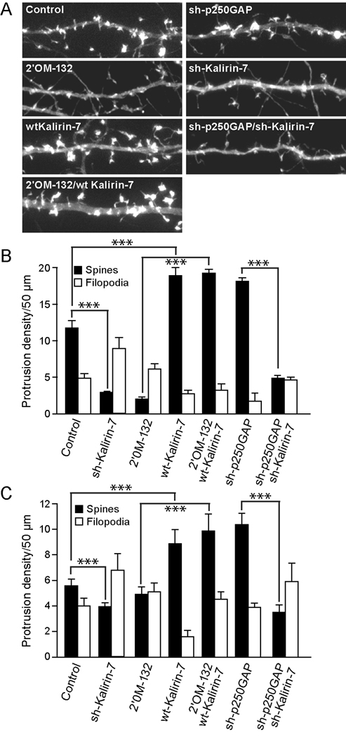
Fig. 5. miR132-regulated spine formation requires the RacGEF, Kalirin-7.
(A, B) Hippocampal neurons DIV 7 were transfected with mRFP-β actin and either empty vector (Control) 2’OM-132, sh-p250GAP, wtKalirin-7, sh-Kalirin-7, or combinations where indicated. On DIV12, the neurons were fixed and imaged. Representative dendrites are shown in A and quantified in B. (C) Hippocampal slices were cultured for 3 days and subjected to biolistic transfection with TFP ± other plasmids as indicated. Slices were allowed to recover for 1 day and then stimulated (where indicated) with 20 µM bicuculline for 2 days. On DIV 6, dendritic protrusions were analyzed for spines and filopodia (± SEM, *** P < 0.001).

