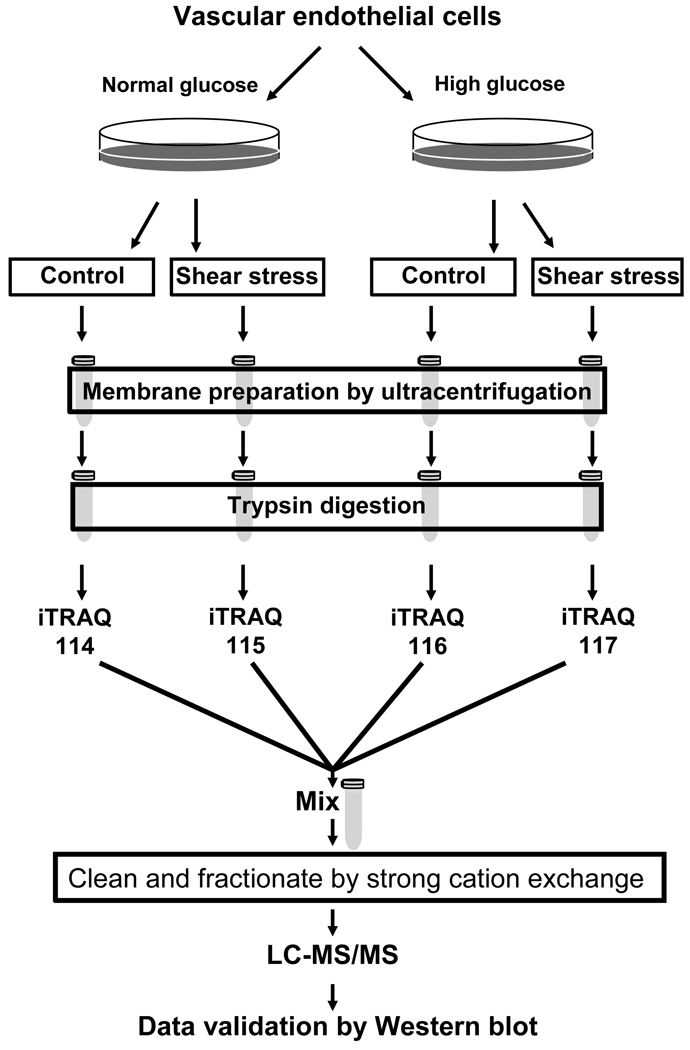
Figure 2. Quantitative proteomic analysis of vascular endothelial cells in high glucose.
Bovine aortic endothelial cells (BAEC) were cultured in normal (5 mM, NG) or high (22 mM, HG) glucose. Cells were either shear stressed or kept in stationary conditions (no flow) as control. Membrane fractions were prepared from the shear stressed and control cells. The membrane associated proteins were digested with trypsin, and the peptides were labeled with iTRAQ reagents. Labeled peptides were combined, fractionated by strong cation exchange chromatography, and analyzed by LC-MS/MS.

