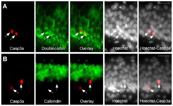
Figure 5. SE-injured DG cells have features of immature neurons.

Caspase-3a-IR DG neurons (arrows) are calbindin-negative and doublecortin-positive. (A) Images of active caspase-3-IR (red), doublecortin-IR (immature neuron marker, green) and Hoechst staining (chromatin dye, white) in DG 24 h following SE. (B) Images of active caspase-3-IR (red), calbindin-IR (mature granule cell marker, green) and Hoechst staining (white) in DG 24 h following SE. Scale bars = 100 μm.
