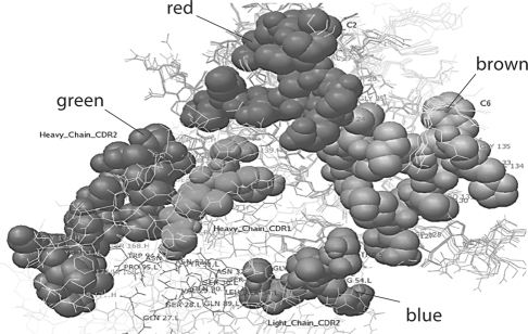
Fig. 5.
Model of human m anti-α3(IV)NC1 F3.1 binding to α3(IV)NC1. F3.1 is shown interacting through heavy chain CDR2 (shown in green) and light chain CDR2 (shown in blue) sites with both the C2 (shown in red) and C6 (shown in brown) epitopes. Note the multiple contact Ab contact regions of the Ab with the repeating epitopes on α3(IV)NC1. The contact regions, however, are different from those of either F1.1. or F2.1.

