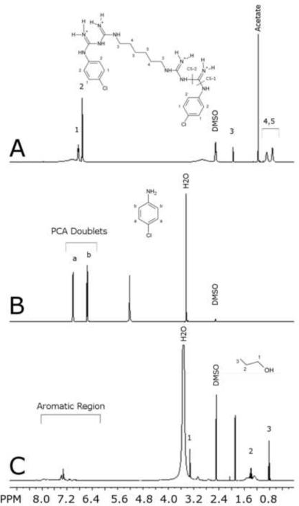
Figure 1.
1H NMR spectrum of (A) chlorhexidine acetate with characteristic peaks at 6.85 ppm and 6.71 ppm (labels 1–5 correspond to labeled chemical structure of chlorhexidine and cleavage sites are labeled CS-1 and CS-2) (B) p-chloroaniline with characteristic peaks at 7.01 ppm and 6.56 ppm (labels a and b correspond to labeled chemical structure of PCA). (C) Reaction precipitate sampled at 60 minutes with n-propanol added at 0.4 mg/ml as an internal standard (labels 1–3 corresponding to labeled chemical structure of n-propanol). All spectra were taken with 400-MHz Varian NMR System at 25°C, acquiring 32 scans, in d6-DMSO solvent.

