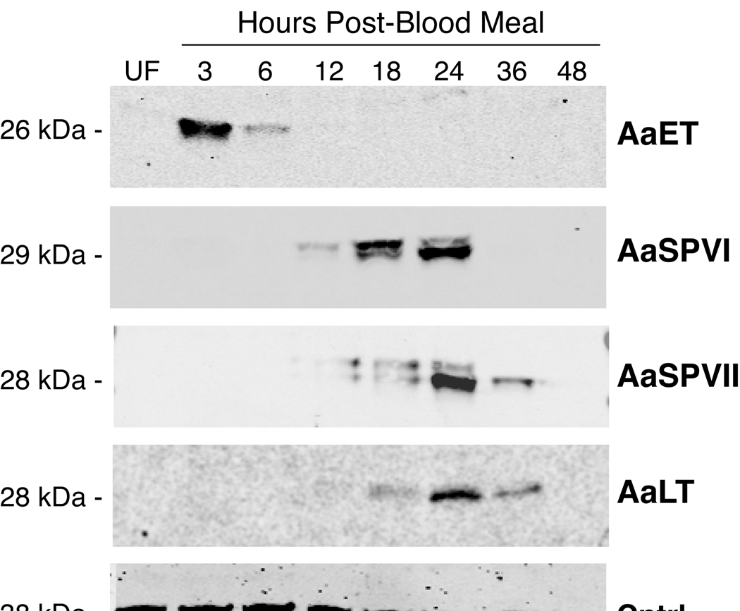
Figure 1.
Representative western blot of serine protease protein expression in the midgut during blood meal digestion. Each lane contains one midgut tissue equivalent of protein extract. Predicted molecular weights for each protein are indicated based on electrophoresed molecular weight standards (not shown) and the number of amino acid residues in the mature form of the serine protease. The serine protease antibody used in each Western blot is listed on the right side, the loading control is a protein detected by an anti-HspBP1 antibody (Raynes et al., 2000).

