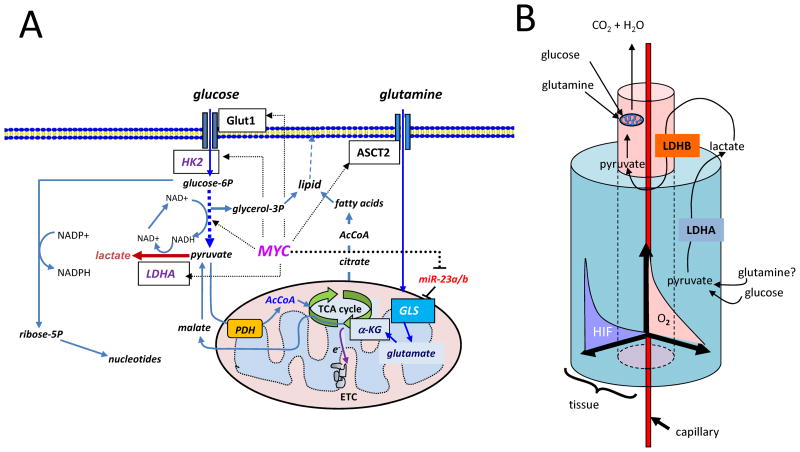
Figure 1.
1A) Diagram depicting glucose and glutamine metabolic pathways and targets (boxed) therein regulated by Myc (dashed arrows). Glucose is transported by Glut1 and phosphorylated by hexokinase 2 (HK2) with subsequent conversion to trioses, producing NADH and ATP, culminating in pyruvate. Intermediate trioses yield glycerol-3-phosphate as a backbone for lipids. Pyruvate can be converted to lactate by lactate dehydrogenase A (LDHA), which is a target of Myc and HIF-1. In the presence of oxygen, pyruvate could be further converted to acetyl-CoA (AcCoA) that is further oxidized in the mitochondria through the tricarboxylic acid (TCA) cycle, which donates high energy electrons (e−) to the electron transport chain (ETC) for the production of ATP and pyrimidine biosynthesis. Citrate transported into cytoplasm from the TCA cycle provides substrate for cytoplasmic acetyl-CoA production, necessary for fatty acid synthesis, which together with glycerol-3-phosphate generate lipids. Glucose-6-phosphate (glucose-6P) can alternatively be catabolized to ribose through the pentose phosphate shunt, which also generates NADPH for redox homeostasis. Glutamine is shown transported into the cell through ASCT2 and converted to glutamate by glutaminase (GLS), which is under the control of MYC through microRNA miR-23a/b. Glutamate is further catabolized to α-ketoglutarate (α-KG) for further oxidation in the TCA cycle. Malate generated from α-ketoglutarate can exit the TCA cycle into the cytoplasm for conversion to pyruvate. 1B) Cartoon depicting a 3-D cutout of a tumor tissue block with a central capillary feeding an inner kernel of cells with oxygen and nutrients. This kernel uses oxidative phosphorylation with glucose and glutamine serving as substrates. As cell proliferate and are pushed away from the blood vessel, an oxygen gradient (O2) is created with a concomitant increase in HIF-1 levels in the peripheral cuff of hypoxic cells, which utilizes glycolysis and perhaps glutaminolysis (conversion of glutamine to lactate). Note that lactate produced by LDHA in the hypoxic cuff is converted to pyruvate by LDHB in the central kernel of cells for oxidation in the mitochondrion (see Figure 1A).

