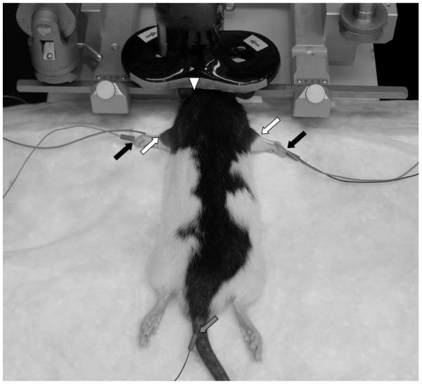
Figure 1. Rat and TMS Coil Setup.
Anesthetized rats were placed into an electrically isolated metal rodent stereotactic frame. EMG was recorded from the brachioradialis muscle bilaterally as the figure-8 TMS coil was positioned with its center anterior and lateral to bregma. Active (white arrow), reference (black arrow) and ground (gray arrow) electrodes are indicated. The TMS coil midline is marked by a white triangle. Note, direction of electrical current (arrows on coil) is constant in with the coil positioned over either the left or the right hemisphere.

